Treated by Dr. Venkat Ram Thyalapalli at Dr Venkatram Thyalapalli
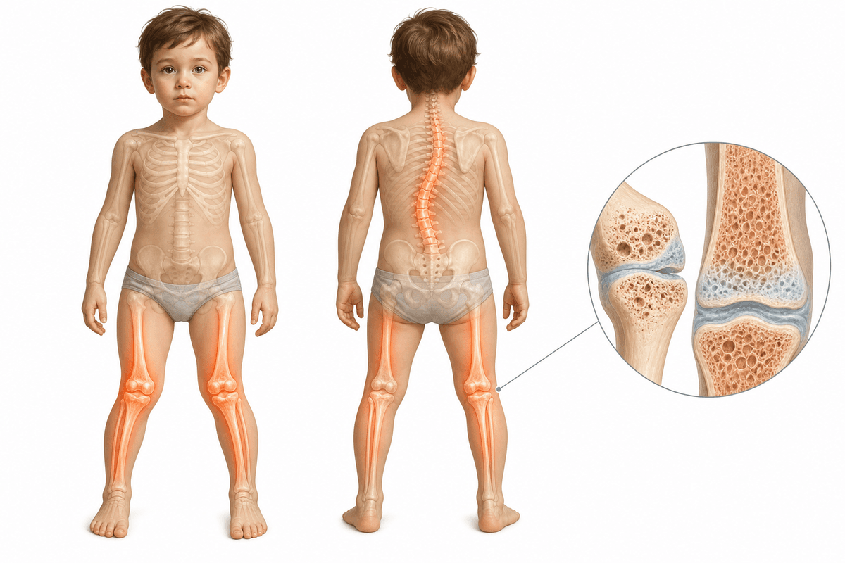
Rickets is a skeletal disorder affecting children in Hyderabad, characterized by soft and weakened bones due to vitamin D, calcium, or phosphate deficiency. The condition leads to bone deformities, delayed growth, and skeletal abnormalities that can significantly impact a child's development. Early detection and comprehensive treatment are essential to prevent permanent deformities. Dr Venkatram Thyalapalli provides specialized pediatric orthopedic care for children with rickets, combining medical management with surgical correction when necessary to restore normal bone development and function.
Most common form caused by inadequate dietary intake of vitamin D, calcium, or insufficient sunlight exposure. Results in defective bone mineralization and characteristic skeletal deformities including bow legs, knock knees, and rachitic rosary.
Rare hereditary disorder affecting vitamin D metabolism. Type 1 involves defective conversion of vitamin D to its active form, while Type 2 involves resistance to vitamin D action at the cellular level, requiring high-dose supplementation.
Genetic disorder characterized by excessive phosphate loss through kidneys, leading to low blood phosphate levels. X-linked hypophosphatemic rickets is the most common inherited form, causing skeletal deformities resistant to standard vitamin D treatment.
Multiple factors can contribute to the development and progression of this condition.
Rickets develops gradually. Recognising symptoms early gives you more treatment options.
From conservative to surgical — we always start with the least invasive option first.
A structured, patient-first approach from first visit to full recovery.
Dr Venkatram Thyalapalli conducts thorough clinical examination assessing skeletal deformities, growth parameters, and developmental milestones. Comprehensive blood work including serum calcium, phosphate, alkaline phosphatase, parathyroid hormone, and vitamin D levels is performed. X-rays of affected bones evaluate the extent of demineralization, metaphyseal changes, and growth plate abnormalities to determine disease severity and guide treatment planning.
Dr Venkatram Thyalapalli develops individualized supplementation protocols based on the specific type and severity of rickets. High-dose vitamin D therapy is initiated with appropriate calcium and phosphate supplementation. Regular monitoring every 4-6 weeks tracks biochemical improvement and bone healing. Nutritional counseling addresses dietary deficiencies, and sunlight exposure recommendations are provided to families.
Dr Venkatram Thyalapalli determines the optimal timing and method for orthopedic correction based on deformity severity, child's age, and response to medical therapy. For mild deformities, bracing protocols are implemented with regular follow-ups. Moderate cases may benefit from guided growth procedures, while severe deformities require comprehensive surgical planning using advanced imaging and computer-assisted correction calculations.
Dr Venkatram Thyalapalli provides ongoing surveillance to ensure continued bone healing, monitor for recurrence, and assess final limb alignment. Regular radiographic follow-ups track bone mineralization and growth plate recovery. Maintenance vitamin D supplementation is prescribed to prevent relapse. Growth monitoring continues until skeletal maturity to identify any residual deformities requiring intervention and ensure optimal functional outcomes.
What to expect at each phase of recovery.
Intensive vitamin D and mineral supplementation begins with close biochemical monitoring. Bone pain typically improves within 2-4 weeks as remineralization starts. Serial X-rays at 6-8 weeks show early healing signs including disappearance of frayed metaphyses and improved bone density. Activity is encouraged as tolerated, and families receive education on nutrition and compliance with supplementation.
Continued medical therapy supports ongoing bone healing and remodeling. Mild to moderate deformities may show spontaneous improvement as bones strengthen, particularly in younger children. Bracing protocols are implemented for persistent angular deformities. Biochemical parameters normalize, and alkaline phosphatase levels decline toward normal ranges. Regular clinical assessments every 2-3 months monitor skeletal alignment and growth progression.
Bone mineralization completes, and skeletal deformities stabilize. Maintenance vitamin D supplementation continues to prevent recurrence. Residual deformities are evaluated for surgical correction if indicated. Children who underwent surgical intervention complete their recovery with restoration of normal limb alignment. Long-term follow-up continues annually until skeletal maturity to ensure sustained correction and monitor for any late complications.
Vitamin D, calcium, phosphate, and alkaline phosphatase levels return to normal ranges within 3-6 months of appropriate supplementation. Parathyroid hormone levels normalize as mineral deficiencies are corrected. Sustained biochemical stability prevents further skeletal deterioration and supports ongoing bone development and mineralization throughout childhood.
Radiographic evidence of bone healing appears within 2-3 months, with complete healing of rachitic changes by 6-12 months. Growth plates normalize, and bone density improves progressively. Early diagnosis and treatment typically result in complete skeletal recovery without residual deformities, particularly in children under 3 years of age.
Muscle strength improves significantly as bone pain resolves and mineral balance is restored. Motor development accelerates with children achieving delayed milestones within months of treatment initiation. Corrected limb alignment allows normal ambulation, running, and age-appropriate physical activities. Most children achieve normal growth velocity and reach genetic height potential with early intervention.
Timely treatment prevents permanent skeletal deformities, short stature, and chronic pain. Dental development normalizes with proper mineralization. Risk of pathological fractures is eliminated once bone strength is restored. Appropriate correction of deformities prevents secondary complications including joint arthritis, abnormal gait patterns, and psychosocial impacts related to appearance.
Untreated rickets leads to progressive skeletal deformities including severe bow legs, knock knees, spinal curvatures, and chest wall abnormalities that become permanent. Children experience chronic bone pain, muscle weakness, short stature, and increased fracture risk. Severe cases can result in respiratory compromise from chest deformities, developmental delays, and lifelong disability requiring complex surgical reconstruction.
Consult Dr Venkatram Thyalapalli immediately if your child shows delayed motor milestones, difficulty walking, visible leg bowing or knock knees, bone pain, muscle weakness, or swelling at wrists and ankles. Children at risk including those with limited sunlight exposure, dark skin, dietary restrictions, or malabsorption disorders should undergo screening. Early evaluation allows prompt treatment initiation, preventing permanent deformities and ensuring optimal skeletal development and lifelong function.
Early treatment means more options and better outcomes. Book a consultation to understand your condition and explore the right path forward.
Consult Venkat Ram Thyalapalli for Rickets at any of these 2 centres — pick the one nearest you.