Treated by Dr. Venkat Ram Thyalapalli at Dr Venkatram Thyalapalli
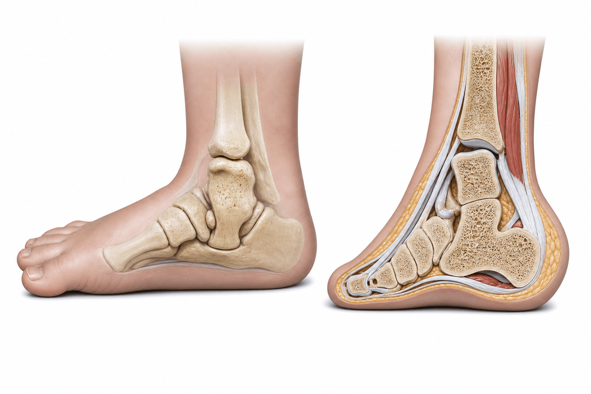
Congenital Vertical Talus is a rare foot deformity present at birth affecting children in Hyderabad and worldwide, characterized by a rigid flatfoot with a rocker-bottom appearance. The condition involves dislocation of the talonavicular joint, where the talus bone is positioned vertically rather than horizontally, causing the foot's abnormal shape and limited flexibility. Dr Venkatram Thyalapalli specializes in the comprehensive diagnosis and surgical correction of this complex pediatric foot deformity.
Occurs as an isolated foot deformity without any associated neuromuscular or genetic conditions. This is the less common form, accounting for approximately 50% of cases, and typically presents with no other systemic abnormalities or syndromes.
Associated with neuromuscular disorders such as myelomeningocele, arthrogryposis, or chromosomal abnormalities like trisomy 18. Approximately 50% of congenital vertical talus cases occur alongside other conditions requiring comprehensive multidisciplinary management.
Affects both feet simultaneously, which is seen in about 50% of all congenital vertical talus cases. Bilateral presentation requires coordinated treatment planning and may indicate higher likelihood of associated syndromic conditions requiring thorough systemic evaluation.
Multiple factors can contribute to the development and progression of this condition.
Congenital Vertical Talus develops gradually. Recognising symptoms early gives you more treatment options.
From conservative to surgical — we always start with the least invasive option first.
A structured, patient-first approach from first visit to full recovery.
Dr Venkatram Thyalapalli performs detailed physical examination of the foot deformity, assessing rigidity, skin condition, and associated neuromuscular findings. Complete medical history is obtained to identify syndromic associations, and advanced imaging including weight-bearing radiographs and ultrasound helps determine the exact position of bones and severity of dislocation.
Dr Thyalapalli develops an individualized treatment protocol based on the child's age, deformity severity, presence of associated conditions, and family circumstances. The treatment plan typically begins with serial manipulation and casting using modified reverse Ponseti principles, with clear timelines for surgical intervention and comprehensive discussion of expected outcomes with parents.
Dr Venkatram Thyalapalli performs meticulous open surgical reduction with extensive experience in pediatric foot reconstruction. The procedure involves careful soft tissue releases, anatomical reduction of the talonavicular joint, temporary pin fixation, and tendon lengthenings, all executed with attention to preserving blood supply and growth plates for optimal long-term foot development.
Dr Thyalapalli provides comprehensive follow-up care including serial cast changes, pin removal at appropriate timing, transition to custom ankle-foot orthoses, and long-term monitoring for recurrence. Parents receive detailed guidance on bracing protocols, physical therapy exercises, and developmental milestone expectations to ensure the best possible functional outcome.
What to expect at each phase of recovery.
First 8-12 weeks after surgery involve strict immobilization in long-leg casts with regular cast changes every 2-3 weeks. Temporary Kirschner wires remain in place for 6-8 weeks to maintain reduction of the talonavicular joint. Close monitoring for swelling, circulation, and cast-related complications is essential during this critical healing period.
Following cast removal at 3 months, custom ankle-foot orthoses are fitted and worn full-time for 6-12 months, then gradually transitioned to nighttime use for several years. Physical therapy begins to restore ankle motion, strengthen muscles, and facilitate age-appropriate motor development. Progressive weight-bearing is encouraged as tolerated with brace support.
Extended follow-up continues until skeletal maturity to monitor for recurrence, foot growth patterns, and functional outcomes. Annual radiographs track bone alignment and joint development. Brace modifications and potential need for revision procedures are assessed. Most children achieve independent walking with good foot position, though some may require orthotic support long-term.
Successful surgical correction achieves a flat, weight-bearing foot position eliminating the rocker-bottom deformity. The foot can make full contact with the ground during standing and walking, allowing for normal shoe wear and improved gait mechanics. This anatomical correction is maintained with appropriate bracing compliance and follow-up care.
Most children achieve independent walking ability by 18-24 months of age following successful treatment. While some residual stiffness may persist compared to normal feet, functional mobility is excellent in the majority of cases. Children can participate in age-appropriate activities including running and sports with minimal limitations.
Corrected foot alignment eliminates abnormal pressure points and allows comfortable shoe fitting. Without the rigid deformity, children experience significantly improved comfort during weight-bearing activities. Long-term pain outcomes are generally favorable when anatomical alignment is achieved and maintained through growth.
Early surgical intervention prevents development of fixed bony deformities, arthritic changes, and compensatory problems in knees, hips, and spine. Proper treatment minimizes the risk of recurrence and reduces the likelihood of requiring extensive reconstructive procedures later in childhood or adolescence.
Untreated congenital vertical talus results in permanent rigid rocker-bottom foot deformity that significantly impairs walking ability and causes severe disability. Children develop abnormal gait patterns, experience difficulty with shoe fitting, and suffer from painful callosities over bony prominences. Progressive secondary deformities develop in the ankles, knees, and hips due to compensatory mechanisms, leading to early-onset arthritis and chronic pain that severely limits quality of life and independence.
Parents should seek immediate evaluation by a pediatric orthopedic specialist like Dr Venkatram Thyalapalli if their newborn has a rigid flatfoot deformity with rocker-bottom appearance that does not correct with gentle manipulation. Early diagnosis within the first few months of life is crucial for optimal treatment outcomes. Any infant with foot deformity, especially if associated with other congenital conditions or neuromuscular disorders, requires prompt specialized assessment to initiate appropriate intervention before the deformity becomes more rigid and difficult to correct.
Early treatment means more options and better outcomes. Book a consultation to understand your condition and explore the right path forward.
Consult Venkat Ram Thyalapalli for Congenital Vertical Talus at any of these 2 centres — pick the one nearest you.