Treated by Dr. Venkat Ram Thyalapalli at Dr Venkatram Thyalapalli
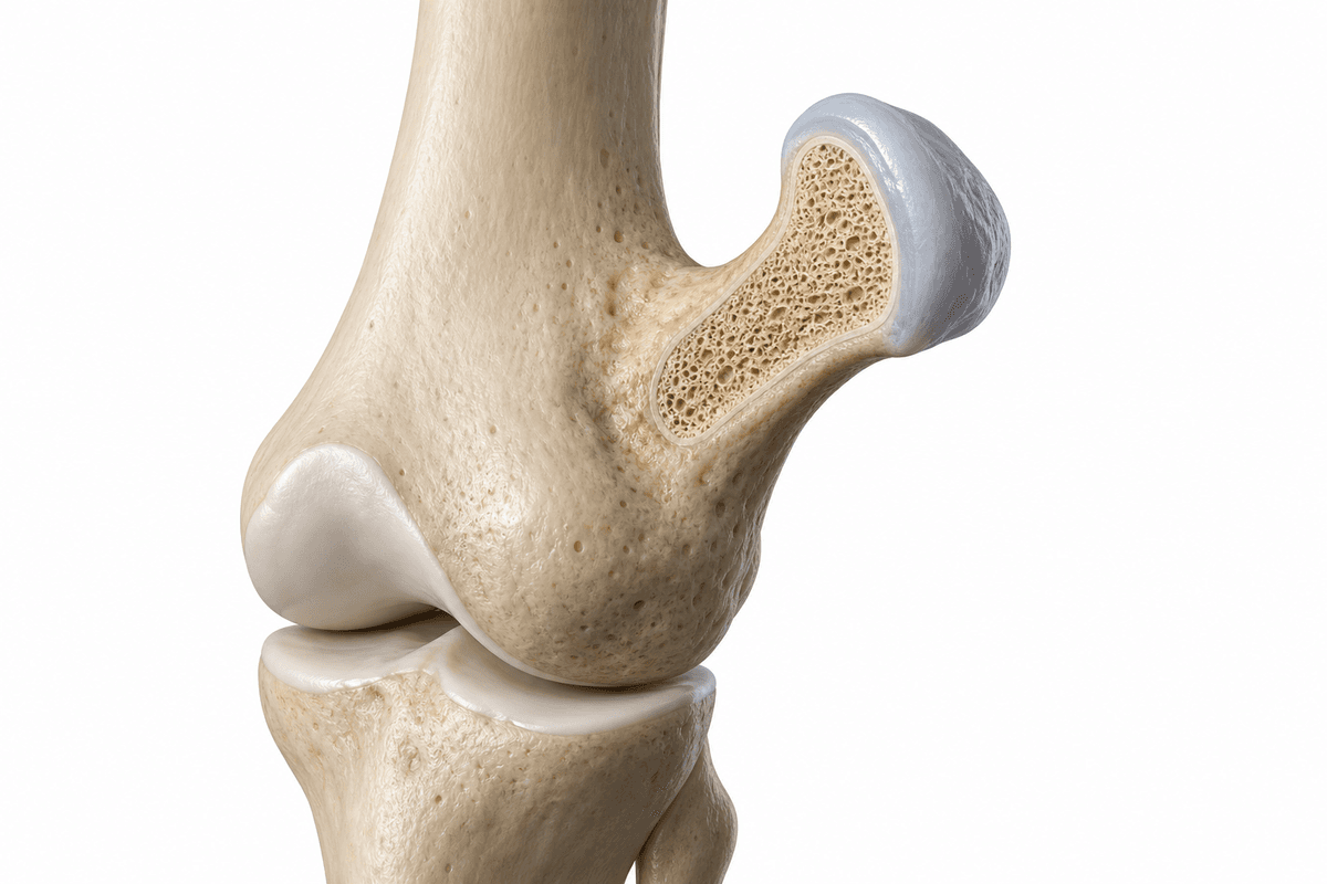
Exostosis, also known as osteochondroma, is a benign bone growth that commonly affects children and adolescents in Hyderabad. These bony projections develop on the surface of bones, typically near growth plates in long bones such as the femur, tibia, or humerus. Dr Venkatram Thyalapalli provides expert diagnosis and surgical treatment for exostosis at his pediatric orthopedic clinic.
A single benign bone growth occurring on one bone, most commonly found near the knee, shoulder, or hip. This is the most frequent type of exostosis, typically discovered during childhood or adolescence when the growth becomes palpable or causes mechanical symptoms.
An inherited genetic condition causing multiple osteochondromas to develop throughout the skeleton. This autosomal dominant disorder affects growth plates and can lead to limb length discrepancies, angular deformities, and requires closer monitoring for potential complications.
A rare type of bone growth that develops beneath or beside the nail bed, most commonly affecting the great toe. This variant causes pain, nail deformity, and local tenderness, requiring specific surgical approach different from long bone exostoses.
Multiple factors can contribute to the development and progression of this condition.
Exostosis develops gradually. Recognising symptoms early gives you more treatment options.
From conservative to surgical — we always start with the least invasive option first.
A structured, patient-first approach from first visit to full recovery.
Dr Venkatram Thyalapalli conducts thorough physical examination to assess the size, location, and characteristics of the exostosis. He evaluates for any neurovascular compromise, joint restriction, or cosmetic concerns while taking detailed medical and family history to identify hereditary patterns.
High-quality X-rays are obtained to visualize the bony growth and its relationship to the underlying bone. When needed, Dr Thyalapalli orders CT scans for three-dimensional assessment or MRI to evaluate the cartilage cap thickness and soft tissue involvement, ensuring accurate surgical planning.
Based on the patient's age, symptoms, growth potential, and imaging findings, Dr Venkatram Thyalapalli develops an individualized treatment strategy. He discusses observation versus surgical options with families, explaining risks, benefits, and expected outcomes to facilitate informed decision-making.
When surgery is indicated, Dr Thyalapalli performs meticulous excision with attention to complete removal while preserving healthy tissue. He provides comprehensive postoperative care including pain management, rehabilitation guidance, and long-term surveillance to monitor for rare recurrence or delayed complications.
What to expect at each phase of recovery.
Initial wound healing occurs with pain managed through prescribed medications. The surgical site is protected with dressings and immobilization if needed. Patients gradually resume light activities while avoiding stress on the operated area, with first follow-up to assess healing.
Physical therapy begins to restore range of motion and strength in the affected limb. Protective restrictions are gradually lifted as bone healing progresses. Most children return to school and non-contact activities during this period, with progressive functional improvement.
Complete return to all activities including sports is typically achieved by 8-12 weeks. Dr Thyalapalli monitors with follow-up X-rays at 3 and 6 months to confirm complete excision and absence of recurrence. Long-term surveillance continues until skeletal maturity, especially for hereditary cases.
Over 95% of patients experience complete relief from pain, mechanical symptoms, and cosmetic concerns following surgical excision. The bony prominence is eliminated, restoring normal contour and function to the affected area with high patient satisfaction.
Removal of exostoses that impinge on joints or restrict motion results in significant improvement in range of movement. Children regain full participation in sports and physical activities without limitations, with normalized biomechanics and movement patterns.
Timely excision prevents potential complications such as neurovascular compression, fracture through the stalk, bursa formation, or malignant transformation. Early intervention in growing children can prevent progressive deformities and growth disturbances.
When complete excision including the cartilage cap is achieved, recurrence rates are very low, typically less than 2% for solitary lesions. Dr Thyalapalli's meticulous surgical technique ensures thorough removal, minimizing the risk of regrowth and need for revision surgery.
Untreated symptomatic exostosis can lead to progressive pain, joint dysfunction, and mechanical complications. Large or strategically located growths may compress adjacent nerves or blood vessels, causing neurological deficits or vascular compromise. In rare cases, particularly with multiple hereditary exostoses, there is a small risk of malignant transformation to chondrosarcoma, necessitating vigilant monitoring and timely intervention when indicated.
Consult Dr Venkatram Thyalapalli if you notice a hard, non-mobile bump on your child's bone, especially near joints, or if an existing growth is enlarging or becoming painful. Immediate evaluation is warranted if there is sudden pain in the exostosis, numbness or tingling in the limb, visible deformity, or restriction in joint movement. Early assessment allows for appropriate monitoring and timely intervention when needed.
Early treatment means more options and better outcomes. Book a consultation to understand your condition and explore the right path forward.
Consult Venkat Ram Thyalapalli for Exostosis at any of these 2 centres — pick the one nearest you.